Balbharti Maharashtra State Board 11th Biology Textbook Solutions
Chapter 16 Skeleton and Movement Textbook Exercise Questions and Answers.
1. Choose the correct option
Question (A).
The functional unit of striated muscle is …………..
a. cross bridges
b. myofibril
c. sarcomere
d. z-band
Answer:
c. sarcomere
Question (B).
A person slips from the staircase and breaks his ankle bone. Which bones are involved?
a. Carpals
b. Tarsal
c. Metacarpals
d. Metatarsals
Answer:
b. Tarsal

Question (C).
Muscle fatigue is due to accumulation of ……..
a. pyruvic acid
b. lactic acid
c. malic acid
d. succinic acid
Answer:
b. lactic acid
Question (D).
Which one of the following is NOT antagonistic muscle pair?
a. Flexo-extensor
b. Adductor-abductor
c. Levator-depressor
d. Sphinetro-suprinater
Answer:
d. Sphinetro-suprinater
Question (E).
Swelling of sprained foot is reduced by soaking in hot water containing a large amount of common salt,
a. due to osmosis
b. due to plasmolysis
c. due to electrolysis
d. due to photolysis
Answer:
a. due to osmosis
Question (F).
Role of calcium in muscle contraction is ……….
a. to break the cross bridges as a cofactor in the hydrolysis of ATP
b. to bind with troponin, changing its shape so that the actin filament is exposed
c. to transmit the action potential across the neuromuscular junction.
d. to re-establish the polarisation of the plasma membrane following an action potential
Answer:
b. to bind with troponin, changing its shape so that the actin filament is exposed
Question (G).
Hyper-secretion of parathormone can cause which of the following disorders?
a. Gout
b. Rheumatoid arthritis
c. Osteoporosis
d. Gull’s disease
Answer:
c. Osteoporosis
Question (H).
Select correct option between two nasal bones

Answer:
(c)
Question 2.
Answer the following questions
Question (A).
What kind of contraction occurs in your neck muscles while you are reading your class assignment?
Answer:
Question (B).
Observe the diagram and enlist importance of ‘A’, ‘B’ and ‘C’.
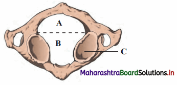
Answer:
Question (C).
Raju intends to train biceps; while exercising using dumbbells, which joints should remain stationary and which should move?
Answer:
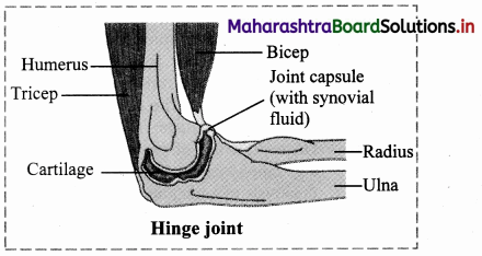
While performing exercise of biceps using dumbbells, the joint which should remain stationary are wrist joint or radiocarpal joint, ball and socket joint of shoulder. The only joint which should move is hinge joint of elbow.
Question (D).
In a road accident, Moses fractured his leg. One of the passers by, tied a wodden plank to the fractured leg while Moses
was rushed to the hospital Was this essential? Why?
Answer:
Question (E).
Sprain is more painful than fracture. Why?
Answer:
[Note: 1st and 2nd degree sprains are not very serious and may be lesser painful than a fracture. Depending on the severity of the injury, intensity of pain will vary.]
Question (F).
Why a red muscle can work for a prolonged period whereas white muscle fibre suffers from fatigue after a shorter work? (Refer to chapter animal tissues.)
Answer:
Thus, red muscle fibres can perform prolonged work and show less fatigue due to accumulation of negligible amount loss or of lactic acid, whereas white muscle fibres suffer from fatigue after a shorter work due to accumulation of higher amount of lactic acid.
3. Answer the following questions in detail
Question (A).
How is the structure of sarcomere suitable for the contractility of the muscle? Explain its function according to sliding
filament theory. (Refer to chapter animal tissues.)
Answer:
i. Sarcomere is the functional unit of myofibril. It has specific arrangement of actin and myosin filaments. The components of sarcomere are organized into variety of bands and zones. Actin and myosin are referred as contractile proteins. Actin is called as thin filament whereas myosin in called as thick filament. The structure of sarcomere:
ii. ‘A’ band – dark bands present at the centre of sarcomere and contain myosin as well as actin.
‘H’ zone or Hensen’s zone – light area present at the centre of ‘A’ band
‘M’ line – present at the centre of ‘H’ zone
‘I’ band – light bands present on the either side of ‘A’ band containing only actin
Z’ line – adjacent ‘I’ bands are separated by ‘Z’ line.
iii. Sliding filament theory: It was put forth by H.E Huxley and A.F Huxley. It is also known as ‘Walk along theory’ or Ratchet theory.
Question (B).
Ragini, a 50 year old office goer, suffered hair-line cracks in her right and left foot in short intervals of time. She was worried about minor jerks leading to hair line cracks in bones. Doctor explained to her why it must be happening and prescribed medicines.
What must be the cause of Ragini’s problem? Why has it occurred? What precautions she should have taken earlier? What care she should take in future?
Answer:
Question (C).
How does structure of actin and myosin help muscle contraction?
Answer:
i. Myosin filament:
ii. Actin filament: It is a complex type of contractile protein. It is made up of three components:
Question (D).
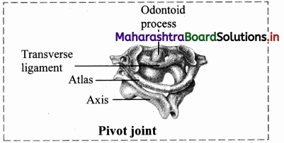
Justify the structure of atlas and axis vertebrae with respect to their position and function.
Answer:
i. Atlas vertebrae:
ii. Axis vertebrae:
Question (E).
Observe the blood report given below and diagnose the possible disorder.
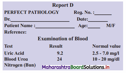
Answer:
On observing Report D, it is clear that the level of uric acid is more than normal, thus the patient must be suffering from gouty arthritis.
Also, the elevated blood urea nitrogen (BUN) indicates dysfunctional liver and/ or kidneys. It generally occurs due to decrease in GFR, caused by renal disease or obstruction of urinary tract.
Question 4.
Write short notes on following points
Question (A).
Actin filament
Answer:
Actin filament: It is a complex type of contractile protein. It is made up of three components:
Question (B).
Myosin filament
Answer:
i. Myosin filament:
Question (C).
Role of calcium ions in contraction and relaxation of muscles.
Answer:
Calcium ions play a major role in contraction and relaxation of muscles.
Question 5.
Draw labelled diagrams
Question (A).
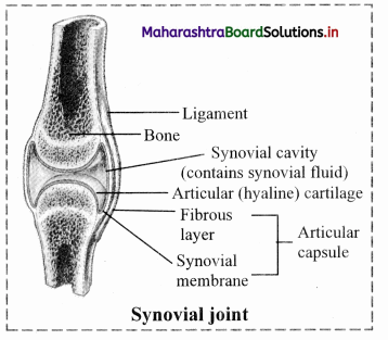
Synovial joint.
Answer:
i. Synovial joints / freely movable joints / diarthroses:
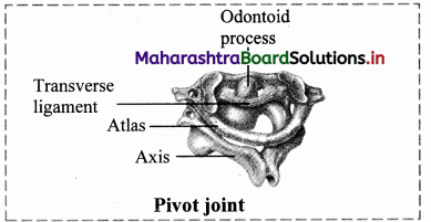
1. Pivot joint: In this type of joint, the rounded or pointed surface of one bone articulates with a ring formed partly by another bone and partly by the ligament. Rotation only around its own longitudinal axis is possible. e.g. in joint between atlas and axis vertebrae, head turns side ways to form ‘NO’ joint.
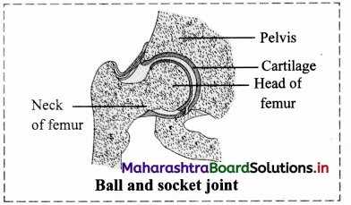
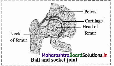
2. Ball and socket joint: The ball like surface of one bone fits into cup like depression of another bone forming a movable joint. Multi-axial movements are possible. This type of joint allows movements along all three axes and in all directions. e.g. Shoulder and hip joint.
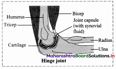
3. Hinge joint: In a hinge joint, convex surface of one bone fits into concave surface of another bone. In most hinge joints one bone remains stationary and other moves. The angular opening and closing motion (like hinge) is possible. In this joint only mono-axial movement takes place like flexion and extension. e.g. Elbow and knee joint.
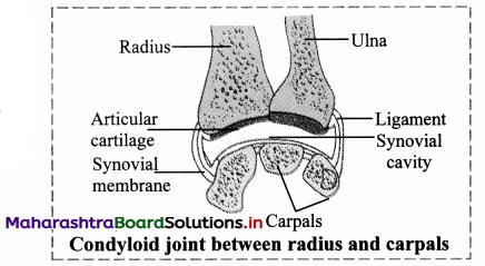
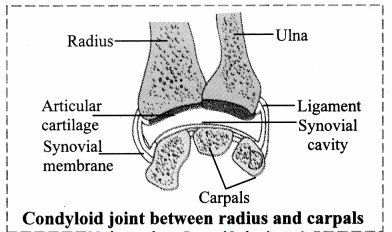
4. Condyloid joint: It is an ellipsoid joint. The convex oval shaped projection of one bone fits into oval shaped depression in another bone. it is a biaxial joint because it permits movement along two axes viz, flexion, extension, abduction, adduction and circumduction is possible. e.g. Metacarpophalangeal joint.
5. Gliding joint: It is a planar joint, where the articulating surfaces of bones are flat or slightly curved. These joints are non-axial because the motion they allow does not occur along an axis or a plane. e.g. Intercarpal and intertarsal joints.
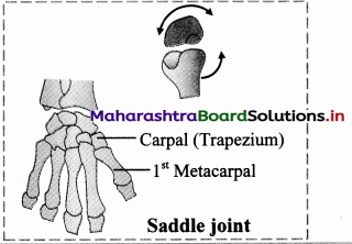
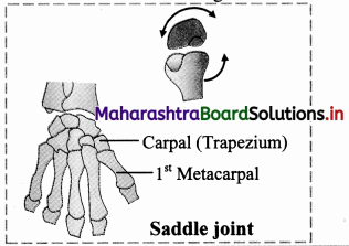
6. Saddle joint: This joint is a characteristic of Homo sapiens. Here the articular surface of one bone is saddle-shaped and that of other bone fits into saddle (each bone forming this joint have both concave and convex areas). It is a modified condyloid joint in which movement is somewhat more free. It is a biaxial joint that allows flexion, extension, abduction, adduction and circumduction.
e.g. Carpometacarpellar joint between carpal (trapezium) and metacarpal of thumb.
Question (B).
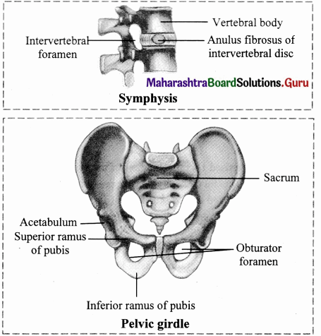
Different cartilagenous joints.
Answer:
Cartilaginous / slightly movable joints / amphiarthroses:
These joints are neither fixed nor freely movable. Articulating bones are held together by hyaline or fibrocartilages. They are further classified as
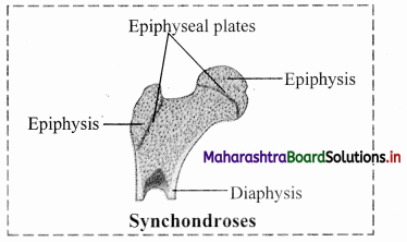
a. Synchondroses: The two bones are held together by hyaline cartilage. They are meant for growth. On completion of growth, the joint gets ossified, e.g. Epiphyseal plate found between epiphysis and diaphysis of a long bone, Rib – Sternum junction.
b. Symphysis: In this type of joint, broad flat disc of fibrocartilage connects two bones. It occurs in mid-line of the body. e.g. Intervertebral discs, manubrium and sternum, pubic symphysis.
Practical / Project :
Identify the following diagrams and demonstrate the concepts in classroom.
Answer:
The diagrams A, B and C represent Class I, Class II and Class III lever respectively.
For description:
[Students are expected to perform the given activity on their own]
12th Biology Digest Chapter 16 Skeleton and Movement Intext Questions and Answers
Movements And Locomotion (Textbook Page No. 193)
Question 1.
Streaming of protoplasm, peristalsis, walking, running, etc. Which of the above-mentioned movements are internal? Which are external? Can you add few more examples?
Answer:
Can you recall? (Textbook Page No. 193)
Question 1.
Which are different types of muscular tissues?
Answer:
Question 2.
Name the type of muscles which bring about running and speaking.
Answer:
Skeletal muscles (Voluntary muscles)
Question 3.
Name the muscles which do not contract as per our will.
Answer:
Involuntary muscles (smooth muscles and cardiac muscles)
Question 4.
Which type of muscles show rhythmic contractions?
Answer:
Cardiac muscles
Question 5.
Which type of muscle is present in the diaphragm of the respiratory system?
Answer:
Skeletal muscle
Question 6.
State the functions of:
Answer:
Can you recall? (Textbook Page No. 193)
Question 1.
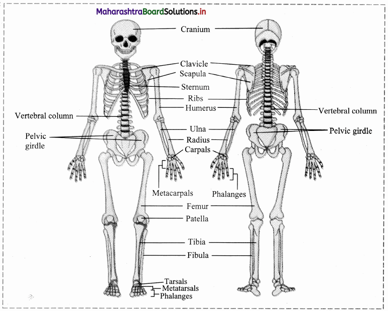
Name the part of human skeleton situated along the vertical axis.
Answer:
Axial skeleton
Question 2.
Give an account of bones of human skull.
Answer:
Skull is made up of 22 bones. It is located at the superior end of vertebral column. The bones of skull are
joined by fixed or immovable joints except for jaw.
Skull consists of cranium or brain box and facial bones.
i. Cranium: It is made up of four median bones and two paired bones.
ii. Facial Bones: Fourteen facial bones give a characteristic shape to the face. The growth of face stops of the
age of 16.
Following bones comprise the facial bones:
Think about it. (Textbook Page No. 193)
Question 1.
Did you ever feel tickling in muscles?
Answer:
Yes, the tickling sensation in muscles can be felt and sometimes it is also accompanied by itching sensation.
Question 2.
What is locomotion?
Answer:
The change in locus of whole body of living organism from one place to another place is called locomotion.
Question 3.
State the four basic types of locomotory movements seen in animals.
Answer:
The four basic types locomotory movements seen in animals are:
Question 4.
Question 1.
Why do muscles show spasm after rigorous contraction?
Answer:
Question 2.
Why do we shiver during winter?
Answer:
Can you tell? (Textbook Page No. 194)
Question 1.
Why are movement and locomotion necessary among animals?
Answer:
Question 2.
All locomotions are movements but all movements are not locomotion. Justify
Answer:
Locomotion occurs when body changes its position, however all movements may not result in locomotion. Thus, all locomotions are movements but all movements are not locomotion.
Question 3.
Kriti was diagnosed with knee tendon injury. She asked the doctor whether she will be able to walk due to the injury? If not then state the reason.
Answer:
Knee tendon injury affects the ability to walk. Kriti may not be able to walk freely as the tendons attached to the bones help in the movement of the parts of skeleton.
Question 4.
What are antagonistic muscles? Explain with example.
Answer:
Question 5.
Describe the antagonistic muscles in detail.
Answer:
Following are the important antagonistic muscles:
Question 6.
Differentiate between:
i. Flexor and extensor muscles
Answer:
ii. Pronator and supinator: Pronator turns the palm downwards and supinator turns the palm upward.
Can you recall? (Textbook Page No. 194)
Question 1.
Comment on contraction of skeletal muscles.
Answer:
Skeletal muscles show quick and strong voluntary contractions. They bring about voluntary movements of the body. For mechanism of muscle contraction:
When the muscles are relaxed, the active sites remain covered with tropomyosin and troponin complex. Due to this, myosin cannot interact with active site of actin and thus contraction cannot occur.
Do You Know How (Textbook Page No. 195)
Question 1.
Do skeletal muscles contract and bring about movement and locomotion?
Answer:
When the muscles are relaxed, the active sites remain covered with tropomyosin and troponin complex. Due to this, myosin cannot interact with active site of actin and thus contraction cannot occur.
Internet my friend. (Textbook Page No. 196)
Question 1.
Collect information about‘T’ tubules of sarcoplasmic reticulum.
Answer:
Can you tell? (Textbook Page No. 197)
Question 1.
Explain the chemical changes taking place in muscle contraction.
Answer:
The muscle undergoes various chemical changes during contraction, they are as follows:
Question 2.
Why are muscle rich in creatine phosphate?
Answer:
Question 3.
Explain the mechanism of muscle contraction and relaxation.
Answer:
Mechanism of muscle contraction:
When the muscles are relaxed, the active sites remain covered with tropomyosin and troponin complex. Due to this, myosin cannot interact with active site of actin and thus contraction cannot occur.
Muscle relaxation: During relaxation, all the events occur in reverse direction as that of muscle contraction.
Question 4.
What do you understand by muscle twitch?
Answer:
Single muscle twitch:
Single muscle twitch: It is a muscle contraction initiated by a single brief-stimulation. It occurs in 3 stages: a latent period of no contraction, a contraction period and a relaxation period.
Do You Know How (Textbook Page No. 198)
Question 1.
Exoskeletal components change from lower to higher group of animals. These include chitinous structures, nails, horns, hooves, scales, hair, shell, plates, fur, muscular foot, tube feet, etc.
Question 1.
Do you know any of these exoskeletal structures help in movement and locomotion?
Answer:
Nails, hooves, scales, plates, muscular foot and tube feet help in movement and locomotion.
Question 2.
How do scales and plates help in movement and locomotion?
Answer:
Scales and plates in reptiles like snakes provide grip to move on rough edgy surfaces.
Question 3.
Are scales of a fish and that of snake similar?
Answer:
Fishes have dermal scales (bony scales), whereas reptiles like snakes have epidermal scales or scutes (horny, tough extensions of outer layer of skin i.e., stratum corneum).
Question 4.
Find out more information about exoskeletal structures and their role in movement and locomotion.
Answer:
Exoskeletal structures: Exoskeleton provide support, help in movement and also provides protection from predators. The exoskeletal structures vary from organism to organism. Echinoderms have tube feet for locomotion whereas molluscs (e.g. Chiton) have muscular foot for movement and locomotion
[Students are expected to find out more information about exoskeletal structures on their own.]
Question 2.
Name the tissues that form the structural framework of the body.
Answer:
Cartilage and bone
Do you remember? (Textbook Page No. 198)
Question 1.
What are the components of skeletal system?
Answer:
The components of skeletal system are bones, tendons, ligaments and joint.
Question 2.
What type of bones are present in our body?
Answer:
Long bones, short bones, flat bones, irregular bones and sesamoid bones.
Question 3.
How do bones help in various ways?
Answer:
Use your brain power. (Textbook Page No. 198)
Question 1.
Can you compare bone, muscle and joint which help in locomotion with any simple machines you have studied earlier?
Answer:
Bone, muscle and joint can be compared to the simple machines called levers. Joints act as fulcrum, respective muscle generates the force required to move the bone associated with joint.
Question 2.
Explain the three types of lever found in human body.
Answer:
The three types of lever are as follows:
Use your brain power. (Textbook Page No. 199)
Question 1.
Why are long bones slightly bent and not straight?
Answer:
Identify and label. (Textbook Page No. 199)
Question 1.
Identify the different bones.
Answer:
Identify and label. (Textbook Page No. 200)
Question 1.
Name A, B, C and D from the given figure and discuss in group.
Answer:
A – Coronal suture,
B – Sagittal suture,
C – Lambdoidal suture,
D – Lateral / Squamous suture
Skull has many sutures (type of immovable joints) present, out of which four prominent ones are:
Can you tell? (Textbook Page No. 201)
Question 1.
Give schematic plan of human skeleton.
Answer:
[Note: Numbers in the bracket indicate the number of bones.]
Can you tell? (Textbook Page No. 201)
Question 1.
Enlist the bones of cranium.
Answer:
Cranium: It is made up of four median bones and two paired bones.
Can you tell? (Textbook Page No. 201)
Question 1.
Write a note on structure and function of skull.
Answer:
i. Structure of skull:
Skull is made up of 22 bones. It is located at the superior end of vertebral column. The bones of skull are joined by fixed or immovable joints except for jaw.
Skull consists of cranium or brain box and facial bones.
i. Cranium: It is made up of four median bones and two paired bones.
ii. Facial Bones: Fourteen facial bones give a characteristic shape to the face. The growth of face stops of the age of 16.
Following bones comprise the facial bones:
ii. Functions of skull:
Internet my friend. (Textbook Page No. 201)
Question 1.
Cleft palate and cleft lip
Answer:
[Students are expected to find more information about Cleft Palate and lip on internet.]
Can you tell? (Textbook Page No. 202)
Question 1.
Why skull is important for us? Enlist few reasons.
Answer:
Functions of skull:
Internet my friend. (Textbook Page No. 202)
Question 1.
Find out information about sinuses present in skull, functions of skull and disorder ‘sinusitis’.
Answer:
Sinuses are the hollow cavities present in the skull. They humidify the air we breathe.
i. The four types of sinuses present in the skull:
ii. Functions of skull:
Functions of skull:
iii. Sinusitis: It is the inflammation of tissue lining the sinuses. Healthy sinuses when get blocked with mucus and germs causing infection which may lead to sinusitis.
[Students are expected to find more information about sinusitis, using the internet.]
Something interesting. (Textbook Page No. 202)
Question 1.
If police suspect strangulation, they carefully inspect hyoid bone and cartilage of larynx. These get fractured during strangulation. V arious such investigations are done in case of suspicious death of an individual where ossification of sutures in skull, width of pelvic girdle, etc. are examined to find out approximate age of victim or gender of victim, etc. You may find out information about forensic science.
Answer:
Forensic science is an application of science which is used in the matter of criminal determination and civil law. It is generally used in investigation of crimes. Forensic scientists collect, preserve and analyze the evidence during the course of investigation.
[Students are expected to find more information about forensic science on internet.]
Try this. (Textbook Page No. 202)
Question 1.
Feel your spine (vertebral column). Is it straight or curved?
Answer:
Our spinc shows four slight curves which are visible when viewed from the sides.
Question 2.
Find information about slipped disc. (Textbook page no.202)
Answer:
If the ligaments of the intervertebral discs become injured, the pressure developed in the nucleus pulposus protrudes posteriorly or into one of the adjacent vertebrae. This is known as slipped disc.
[Students are expected to find more information using the internet.]
Can you tell? (Textbook Page No. 204)
Question 1.
Write a note on curvatures of vertebral column and mention their importance.
Answer:
Question 2.
Explain the structure of typical vertebra.
Answer:
Question 2.
How will you identify a thoracic vertebra?
Answer:
Thoracic vertebrae can be identified on the basis of centrum, as the centrum of the thoracic vertebrae is heart shaped.
Can you recall? (Textbook Page No. 206)
Question 1.
How does humerus form ball and socket joint? Where is it located?
Answer:
The head of humerus fits into the glenoid cavity of scapula and forms ball and socket joint. it is located in shoulder and hips.
Can you tell? (Textbook Page No. 208)
Question 1.
Differentiate between the skeleton of palm and foot.
Answer:
Question 2.
Explain the longest bone in human body.
Answer:
Femur: The thigh bone is the longest bone in the body. The head is joined to shaft at an angle by a short neck. It forms ball and socket joint with acetabulum cavity of coxal bone. The lower one third region of shaft is triangular flattened area called popliteal surface. Distal end has two condyles that articulate with tibia and fibula.
Internet my friend. (Textbook Page No. 212)
Question 1.
Find out information about types of fractures and how they heal.
Answer:
[Source: Tortora, G., Derrickson, B. Principles of Anatomy and Physiology. 15th Edition]
[Students are expected to find out more information about healing of fractures using the internet.]
Do you remember? (Textbook Page No. 208)
Question 1.
What are joints? What are the types?
Answer:
i. A point where two or more bones get articulated is called joint or articulation or athrosis.
They are classified based on degree of flexibility or movement they permit into lastly synovial or freely movable or diarthroses type of joints.
ii. Synarthroses / fibrous joints / movable joints:
In this joint, the articulating bones are held together by means of fibrous connective tissue. Bones do not exhibit movement. Hence, it is immovable or fixed type of joint. Synarthroses are further classified into sutures, syndesmoses and gomphoses.
iii. Cartilaginous / slightly movable joints / amphiarthroses:
These joints are neither fixed nor freely movable. Articulating bones are held together by hyaline or
fibrocartilages. They are further classified as
iv. Synovial joints / freely movable joints / diarthroses:
g. The types of synovial joints are on follows:
1. Pivot joint: In this type of joint, the rounded or pointed surface of one bone articulates with a ring formed partly by another bone and partly by the ligament. Rotation only around its own longitudinal axis is possible. e.g. in joint between atlas and axis vertebrae, head turns side ways to form ‘NO’ joint.
2. BaIl and socket joint: The ball like surface of one bone fits into cup like depression of another bone forming a movable joint. Multi-axial movements are possible. This type of joint allows movements along all three axes and in all directions. e.g. Shoulder and hip joint.
3. Hinge joint: In a hinge joint, convex surface of one bone fits into concave surface of another bone. In most hinge joints one bone remains stationary and other moves. The angular opening and closing motion (like hinge) is possible. In this joint only mono-axial movement takes place like flexion and extension. e.g. Elbow and knee joint.
4. Condyloid joint: It is an ellipsoid joint. The convex oval shaped projection of one bone fits into oval shaped depression in another bone. It is a biaxial joint because it permits movement along two axes viz, flexion, extension, abduction, adduction and circumduction is possible. e.g. Metacarpophalangeal joint.
5. Gliding joint: It is a planar joint, where the articulating surfaces of bones are flat or slightly curved. These joints are non-axial because the motion they allow does not occur along an axis or a plane. e.g. Intercarpal and intertarsal joints.
Saddle joint: This joint is a characteristic of Homo sapiens. Here the articular surface of one bone is saddle-shaped and that of other bone fits into saddle (each bone forming this joint have both concave and convex areas). It is a modified condyloid joint in which movement is somewhat more free. It is a biaxial joint that allows flexion, extension, abduction, adduction and circumduction. e.g. Carpometacarpellar joint between carpal (trapezium) and metacarpal of thumb.
Imagine. (Textbook Page No. 208)
Question 1.
If your elbow joint would be a fixed type of joint and joint between teeth and gum would be freely movable.
Answer:
Use your brain power. (Textbook Page No. 210)
Question 1.
Why are warming up rounds essential before regular exercise?
Answer:
Can you tell? (Textbook Page No. 211)
Question 1.
Classify various types of joints found in human body. Present the information in the form of chart. Give example of each type.
Answer:
i. A point where two or more bones get articulated is called joint or articulation or athrosis.
They are classified based on degree of flexibility or movement they permit into lastly synovial or freely movable or diarthroses type of joints.
ii. Synarthroses / fibrous joints / movable joints:
In this joint, the articulating bones are held together by means of fibrous connective tissue. Bones do not exhibit movement. Hence, it is immovable or fixed type of joint. Synarthroses are further classified into sutures, syndesmoses and gomphoses.
iii. Cartilaginous / slightly movable joints / amphiarthroses:
These joints are neither fixed nor freely movable. Articulating bones are held together by hyaline or
fibrocartilages. They are further classified as
iv. Synovial joints / freely movable joints / diarthroses:
g. The types of synovial joints are on follows:
1. Pivot joint: In this type of joint, the rounded or pointed surface of one bone articulates with a ring formed partly by another bone and partly by the ligament. Rotation only around its own longitudinal axis is possible. e.g. in joint between atlas and axis vertebrae, head turns side ways to form ‘NO’ joint.
2. BaIl and socket joint: The ball like surface of one bone fits into cup like depression of another bone forming a movable joint. Multi-axial movements are possible. This type of joint allows movements along all three axes and in all directions. e.g. Shoulder and hip joint.
3. Hinge joint: In a hinge joint, convex surface of one bone fits into concave surface of another bone. In most hinge joints one bone remains stationary and other moves. The angular opening and closing motion (like hinge) is possible. In this joint only mono-axial movement takes place like flexion and extension. e.g. Elbow and knee joint.
4. Condyloid joint: It is an ellipsoid joint. The convex oval shaped projection of one bone fits into oval shaped depression in another bone. It is a biaxial joint because it permits movement along two axes viz, flexion, extension, abduction, adduction and circumduction is possible. e.g. Metacarpophalangeal joint.
5. Gliding joint: It is a planar joint, where the articulating surfaces of bones are flat or slightly curved. These joints are non-axial because the motion they allow does not occur along an axis or a plane. e.g. Intercarpal and intertarsal joints.
Saddle joint: This joint is a characteristic of Homo sapiens. Here the articular surface of one bone is saddle-shaped and that of other bone fits into saddle (each bone forming this joint have both concave and convex areas). It is a modified condyloid joint in which movement is somewhat more free. It is a biaxial joint that allows flexion, extension, abduction, adduction and circumduction. e.g. Carpometacarpellar joint between carpal (trapezium) and metacarpal of thumb.
[Students are expected to prepare a chart on their own.]
Can you tell? (Textbook Page No. 211)
Question 1.
Human beings can hold an object in a better manner than monkeys. Why?
Answer:
[Note: Gorillas, chimpanzees, orangutans and some other variants of apes have opposable thumb.]
Internet my friend. (Textbook Page No. 211)
Question 92.
Now a days we hear from many elderly people that they are undergoing knee replacement surgery. Find out why one has to undergo knee replacement; how it is carried out and how it can be prevented.
Answer:
Knee replacement is done in following cases:
[Students are expected to find out more information about knee replacement on internet]
Find out. (Textbook Page No. 212)
Question 1.
You must have heard of Sachin Tendulkar suffering from ‘tennis elbow’, a cricketer suffering from a disorder named after another game. Can common people too suffer from this disorder? Find out more information about this disorder.
Answer:
[Students are expected to find more information about tennis elbow on their own.]
Chapter 16 Skeleton and Movement Textbook Exercise Questions and Answers.
1. Choose the correct option
Question (A).
The functional unit of striated muscle is …………..
a. cross bridges
b. myofibril
c. sarcomere
d. z-band
Answer:
c. sarcomere
Question (B).
A person slips from the staircase and breaks his ankle bone. Which bones are involved?
a. Carpals
b. Tarsal
c. Metacarpals
d. Metatarsals
Answer:
b. Tarsal
Question (C).
Muscle fatigue is due to accumulation of ……..
a. pyruvic acid
b. lactic acid
c. malic acid
d. succinic acid
Answer:
b. lactic acid
Question (D).
Which one of the following is NOT antagonistic muscle pair?
a. Flexo-extensor
b. Adductor-abductor
c. Levator-depressor
d. Sphinetro-suprinater
Answer:
d. Sphinetro-suprinater
Question (E).
Swelling of sprained foot is reduced by soaking in hot water containing a large amount of common salt,
a. due to osmosis
b. due to plasmolysis
c. due to electrolysis
d. due to photolysis
Answer:
a. due to osmosis
Question (F).
Role of calcium in muscle contraction is ……….
a. to break the cross bridges as a cofactor in the hydrolysis of ATP
b. to bind with troponin, changing its shape so that the actin filament is exposed
c. to transmit the action potential across the neuromuscular junction.
d. to re-establish the polarisation of the plasma membrane following an action potential
Answer:
b. to bind with troponin, changing its shape so that the actin filament is exposed
Question (G).
Hyper-secretion of parathormone can cause which of the following disorders?
a. Gout
b. Rheumatoid arthritis
c. Osteoporosis
d. Gull’s disease
Answer:
c. Osteoporosis
Question (H).
Select correct option between two nasal bones
Answer:
(c)
Question 2.
Answer the following questions
Question (A).
What kind of contraction occurs in your neck muscles while you are reading your class assignment?
Answer:
Question (B).
Observe the diagram and enlist importance of ‘A’, ‘B’ and ‘C’.
Answer:
Question (C).
Raju intends to train biceps; while exercising using dumbbells, which joints should remain stationary and which should move?
Answer:
While performing exercise of biceps using dumbbells, the joint which should remain stationary are wrist joint or radiocarpal joint, ball and socket joint of shoulder. The only joint which should move is hinge joint of elbow.
Question (D).
In a road accident, Moses fractured his leg. One of the passers by, tied a wodden plank to the fractured leg while Moses
was rushed to the hospital Was this essential? Why?
Answer:
Question (E).
Sprain is more painful than fracture. Why?
Answer:
[Note: 1st and 2nd degree sprains are not very serious and may be lesser painful than a fracture. Depending on the severity of the injury, intensity of pain will vary.]
Question (F).
Why a red muscle can work for a prolonged period whereas white muscle fibre suffers from fatigue after a shorter work? (Refer to chapter animal tissues.)
Answer:
Thus, red muscle fibres can perform prolonged work and show less fatigue due to accumulation of negligible amount loss or of lactic acid, whereas white muscle fibres suffer from fatigue after a shorter work due to accumulation of higher amount of lactic acid.
3. Answer the following questions in detail
Question (A).
How is the structure of sarcomere suitable for the contractility of the muscle? Explain its function according to sliding
filament theory. (Refer to chapter animal tissues.)
Answer:
i. Sarcomere is the functional unit of myofibril. It has specific arrangement of actin and myosin filaments. The components of sarcomere are organized into variety of bands and zones. Actin and myosin are referred as contractile proteins. Actin is called as thin filament whereas myosin in called as thick filament. The structure of sarcomere:
ii. ‘A’ band – dark bands present at the centre of sarcomere and contain myosin as well as actin.
‘H’ zone or Hensen’s zone – light area present at the centre of ‘A’ band
‘M’ line – present at the centre of ‘H’ zone
‘I’ band – light bands present on the either side of ‘A’ band containing only actin
Z’ line – adjacent ‘I’ bands are separated by ‘Z’ line.
iii. Sliding filament theory: It was put forth by H.E Huxley and A.F Huxley. It is also known as ‘Walk along theory’ or Ratchet theory.
Question (B).
Ragini, a 50 year old office goer, suffered hair-line cracks in her right and left foot in short intervals of time. She was worried about minor jerks leading to hair line cracks in bones. Doctor explained to her why it must be happening and prescribed medicines.
What must be the cause of Ragini’s problem? Why has it occurred? What precautions she should have taken earlier? What care she should take in future?
Answer:
Question (C).
How does structure of actin and myosin help muscle contraction?
Answer:
i. Myosin filament:
ii. Actin filament: It is a complex type of contractile protein. It is made up of three components:
Question (D).
Justify the structure of atlas and axis vertebrae with respect to their position and function.
Answer:
i. Atlas vertebrae:
ii. Axis vertebrae:
Question (E).
Observe the blood report given below and diagnose the possible disorder.
Answer:
On observing Report D, it is clear that the level of uric acid is more than normal, thus the patient must be suffering from gouty arthritis.
Also, the elevated blood urea nitrogen (BUN) indicates dysfunctional liver and/ or kidneys. It generally occurs due to decrease in GFR, caused by renal disease or obstruction of urinary tract.
Question 4.
Write short notes on following points
Question (A).
Actin filament
Answer:
Actin filament: It is a complex type of contractile protein. It is made up of three components:
Question (B).
Myosin filament
Answer:
i. Myosin filament:
Question (C).
Role of calcium ions in contraction and relaxation of muscles.
Answer:
Calcium ions play a major role in contraction and relaxation of muscles.
Question 5.
Draw labelled diagrams
Question (A).
Synovial joint.
Answer:
i. Synovial joints / freely movable joints / diarthroses:
1. Pivot joint: In this type of joint, the rounded or pointed surface of one bone articulates with a ring formed partly by another bone and partly by the ligament. Rotation only around its own longitudinal axis is possible. e.g. in joint between atlas and axis vertebrae, head turns side ways to form ‘NO’ joint.
2. Ball and socket joint: The ball like surface of one bone fits into cup like depression of another bone forming a movable joint. Multi-axial movements are possible. This type of joint allows movements along all three axes and in all directions. e.g. Shoulder and hip joint.
3. Hinge joint: In a hinge joint, convex surface of one bone fits into concave surface of another bone. In most hinge joints one bone remains stationary and other moves. The angular opening and closing motion (like hinge) is possible. In this joint only mono-axial movement takes place like flexion and extension. e.g. Elbow and knee joint.
4. Condyloid joint: It is an ellipsoid joint. The convex oval shaped projection of one bone fits into oval shaped depression in another bone. it is a biaxial joint because it permits movement along two axes viz, flexion, extension, abduction, adduction and circumduction is possible. e.g. Metacarpophalangeal joint.
5. Gliding joint: It is a planar joint, where the articulating surfaces of bones are flat or slightly curved. These joints are non-axial because the motion they allow does not occur along an axis or a plane. e.g. Intercarpal and intertarsal joints.
6. Saddle joint: This joint is a characteristic of Homo sapiens. Here the articular surface of one bone is saddle-shaped and that of other bone fits into saddle (each bone forming this joint have both concave and convex areas). It is a modified condyloid joint in which movement is somewhat more free. It is a biaxial joint that allows flexion, extension, abduction, adduction and circumduction.
e.g. Carpometacarpellar joint between carpal (trapezium) and metacarpal of thumb.
Question (B).
Different cartilagenous joints.
Answer:
Cartilaginous / slightly movable joints / amphiarthroses:
These joints are neither fixed nor freely movable. Articulating bones are held together by hyaline or fibrocartilages. They are further classified as
a. Synchondroses: The two bones are held together by hyaline cartilage. They are meant for growth. On completion of growth, the joint gets ossified, e.g. Epiphyseal plate found between epiphysis and diaphysis of a long bone, Rib – Sternum junction.
b. Symphysis: In this type of joint, broad flat disc of fibrocartilage connects two bones. It occurs in mid-line of the body. e.g. Intervertebral discs, manubrium and sternum, pubic symphysis.
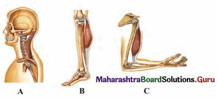
Practical / Project :
Identify the following diagrams and demonstrate the concepts in classroom.
Answer:
The diagrams A, B and C represent Class I, Class II and Class III lever respectively.
For description:
[Students are expected to perform the given activity on their own]
12th Biology Digest Chapter 16 Skeleton and Movement Intext Questions and Answers
Movements And Locomotion (Textbook Page No. 193)
Question 1.
Streaming of protoplasm, peristalsis, walking, running, etc. Which of the above-mentioned movements are internal? Which are external? Can you add few more examples?
Answer:
Can you recall? (Textbook Page No. 193)
Question 1.
Which are different types of muscular tissues?
Answer:
Question 2.
Name the type of muscles which bring about running and speaking.
Answer:
Skeletal muscles (Voluntary muscles)
Question 3.
Name the muscles which do not contract as per our will.
Answer:
Involuntary muscles (smooth muscles and cardiac muscles)
Question 4.
Which type of muscles show rhythmic contractions?
Answer:
Cardiac muscles
Question 5.
Which type of muscle is present in the diaphragm of the respiratory system?
Answer:
Skeletal muscle
Question 6.
State the functions of:
Answer:
Can you recall? (Textbook Page No. 193)
Question 1.
Name the part of human skeleton situated along the vertical axis.
Answer:
Axial skeleton
Question 2.
Give an account of bones of human skull.
Answer:
Skull is made up of 22 bones. It is located at the superior end of vertebral column. The bones of skull are
joined by fixed or immovable joints except for jaw.
Skull consists of cranium or brain box and facial bones.
i. Cranium: It is made up of four median bones and two paired bones.
ii. Facial Bones: Fourteen facial bones give a characteristic shape to the face. The growth of face stops of the
age of 16.
Following bones comprise the facial bones:
Think about it. (Textbook Page No. 193)
Question 1.
Did you ever feel tickling in muscles?
Answer:
Yes, the tickling sensation in muscles can be felt and sometimes it is also accompanied by itching sensation.
Question 2.
What is locomotion?
Answer:
The change in locus of whole body of living organism from one place to another place is called locomotion.
Question 3.
State the four basic types of locomotory movements seen in animals.
Answer:
The four basic types locomotory movements seen in animals are:
Question 4.
Question 1.
Why do muscles show spasm after rigorous contraction?
Answer:
Question 2.
Why do we shiver during winter?
Answer:
Can you tell? (Textbook Page No. 194)
Question 1.
Why are movement and locomotion necessary among animals?
Answer:
Question 2.
All locomotions are movements but all movements are not locomotion. Justify
Answer:
Locomotion occurs when body changes its position, however all movements may not result in locomotion. Thus, all locomotions are movements but all movements are not locomotion.
Question 3.
Kriti was diagnosed with knee tendon injury. She asked the doctor whether she will be able to walk due to the injury? If not then state the reason.
Answer:
Knee tendon injury affects the ability to walk. Kriti may not be able to walk freely as the tendons attached to the bones help in the movement of the parts of skeleton.
Question 4.
What are antagonistic muscles? Explain with example.
Answer:
Question 5.
Describe the antagonistic muscles in detail.
Answer:
Following are the important antagonistic muscles:
Question 6.
Differentiate between:
i. Flexor and extensor muscles
Answer:
| Flexor Muscles | Extensor Muscles | ||
| a. | Flexor muscles contract and bring about the bending or flexion of joint. | a. | Extensor muscles contract and bring about the straightening or extension of joint. |
| b. | These muscles decrease the angle between the bones on two sides of a joint. | b. | These muscles increase the angle between the components of limb. |
| e.g. | Biceps | e.g. | Triceps |
Can you recall? (Textbook Page No. 194)
Question 1.
Comment on contraction of skeletal muscles.
Answer:
Skeletal muscles show quick and strong voluntary contractions. They bring about voluntary movements of the body. For mechanism of muscle contraction:
When the muscles are relaxed, the active sites remain covered with tropomyosin and troponin complex. Due to this, myosin cannot interact with active site of actin and thus contraction cannot occur.
Do You Know How (Textbook Page No. 195)
Question 1.
Do skeletal muscles contract and bring about movement and locomotion?
Answer:
When the muscles are relaxed, the active sites remain covered with tropomyosin and troponin complex. Due to this, myosin cannot interact with active site of actin and thus contraction cannot occur.
Internet my friend. (Textbook Page No. 196)
Question 1.
Collect information about‘T’ tubules of sarcoplasmic reticulum.
Answer:
Can you tell? (Textbook Page No. 197)
Question 1.
Explain the chemical changes taking place in muscle contraction.
Answer:
The muscle undergoes various chemical changes during contraction, they are as follows:
Question 2.
Why are muscle rich in creatine phosphate?
Answer:
Question 3.
Explain the mechanism of muscle contraction and relaxation.
Answer:
Mechanism of muscle contraction:
When the muscles are relaxed, the active sites remain covered with tropomyosin and troponin complex. Due to this, myosin cannot interact with active site of actin and thus contraction cannot occur.
Muscle relaxation: During relaxation, all the events occur in reverse direction as that of muscle contraction.
Question 4.
What do you understand by muscle twitch?
Answer:
Single muscle twitch:
Single muscle twitch: It is a muscle contraction initiated by a single brief-stimulation. It occurs in 3 stages: a latent period of no contraction, a contraction period and a relaxation period.
Do You Know How (Textbook Page No. 198)
Question 1.
Exoskeletal components change from lower to higher group of animals. These include chitinous structures, nails, horns, hooves, scales, hair, shell, plates, fur, muscular foot, tube feet, etc.
Question 1.
Do you know any of these exoskeletal structures help in movement and locomotion?
Answer:
Nails, hooves, scales, plates, muscular foot and tube feet help in movement and locomotion.
Question 2.
How do scales and plates help in movement and locomotion?
Answer:
Scales and plates in reptiles like snakes provide grip to move on rough edgy surfaces.
Question 3.
Are scales of a fish and that of snake similar?
Answer:
Fishes have dermal scales (bony scales), whereas reptiles like snakes have epidermal scales or scutes (horny, tough extensions of outer layer of skin i.e., stratum corneum).
Question 4.
Find out more information about exoskeletal structures and their role in movement and locomotion.
Answer:
Exoskeletal structures: Exoskeleton provide support, help in movement and also provides protection from predators. The exoskeletal structures vary from organism to organism. Echinoderms have tube feet for locomotion whereas molluscs (e.g. Chiton) have muscular foot for movement and locomotion
[Students are expected to find out more information about exoskeletal structures on their own.]
Question 2.
Name the tissues that form the structural framework of the body.
Answer:
Cartilage and bone
Do you remember? (Textbook Page No. 198)
Question 1.
What are the components of skeletal system?
Answer:
The components of skeletal system are bones, tendons, ligaments and joint.
Question 2.
What type of bones are present in our body?
Answer:
Long bones, short bones, flat bones, irregular bones and sesamoid bones.
Question 3.
How do bones help in various ways?
Answer:
Use your brain power. (Textbook Page No. 198)
Question 1.
Can you compare bone, muscle and joint which help in locomotion with any simple machines you have studied earlier?
Answer:
Bone, muscle and joint can be compared to the simple machines called levers. Joints act as fulcrum, respective muscle generates the force required to move the bone associated with joint.
Question 2.
Explain the three types of lever found in human body.
Answer:
The three types of lever are as follows:
Use your brain power. (Textbook Page No. 199)
Question 1.
Why are long bones slightly bent and not straight?
Answer:
Identify and label. (Textbook Page No. 199)
Question 1.
Identify the different bones.
Answer:
Identify and label. (Textbook Page No. 200)
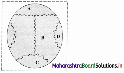
Question 1.
Name A, B, C and D from the given figure and discuss in group.
Answer:
A – Coronal suture,
B – Sagittal suture,
C – Lambdoidal suture,
D – Lateral / Squamous suture
Skull has many sutures (type of immovable joints) present, out of which four prominent ones are:
Can you tell? (Textbook Page No. 201)
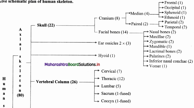
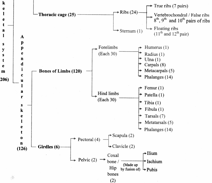
Question 1.
Give schematic plan of human skeleton.
Answer:
[Note: Numbers in the bracket indicate the number of bones.]
Can you tell? (Textbook Page No. 201)
Question 1.
Enlist the bones of cranium.
Answer:
Cranium: It is made up of four median bones and two paired bones.
Can you tell? (Textbook Page No. 201)
Question 1.
Write a note on structure and function of skull.
Answer:
i. Structure of skull:
Skull is made up of 22 bones. It is located at the superior end of vertebral column. The bones of skull are joined by fixed or immovable joints except for jaw.
Skull consists of cranium or brain box and facial bones.
i. Cranium: It is made up of four median bones and two paired bones.
ii. Facial Bones: Fourteen facial bones give a characteristic shape to the face. The growth of face stops of the age of 16.
Following bones comprise the facial bones:
ii. Functions of skull:
Internet my friend. (Textbook Page No. 201)
Question 1.
Cleft palate and cleft lip
Answer:
[Students are expected to find more information about Cleft Palate and lip on internet.]
Can you tell? (Textbook Page No. 202)
Question 1.
Why skull is important for us? Enlist few reasons.
Answer:
Functions of skull:
Internet my friend. (Textbook Page No. 202)
Question 1.
Find out information about sinuses present in skull, functions of skull and disorder ‘sinusitis’.
Answer:
Sinuses are the hollow cavities present in the skull. They humidify the air we breathe.
i. The four types of sinuses present in the skull:
ii. Functions of skull:
Functions of skull:
iii. Sinusitis: It is the inflammation of tissue lining the sinuses. Healthy sinuses when get blocked with mucus and germs causing infection which may lead to sinusitis.
[Students are expected to find more information about sinusitis, using the internet.]
Something interesting. (Textbook Page No. 202)
Question 1.
If police suspect strangulation, they carefully inspect hyoid bone and cartilage of larynx. These get fractured during strangulation. V arious such investigations are done in case of suspicious death of an individual where ossification of sutures in skull, width of pelvic girdle, etc. are examined to find out approximate age of victim or gender of victim, etc. You may find out information about forensic science.
Answer:
Forensic science is an application of science which is used in the matter of criminal determination and civil law. It is generally used in investigation of crimes. Forensic scientists collect, preserve and analyze the evidence during the course of investigation.
[Students are expected to find more information about forensic science on internet.]
Try this. (Textbook Page No. 202)
Question 1.
Feel your spine (vertebral column). Is it straight or curved?
Answer:
Our spinc shows four slight curves which are visible when viewed from the sides.
Question 2.
Find information about slipped disc. (Textbook page no.202)
Answer:
If the ligaments of the intervertebral discs become injured, the pressure developed in the nucleus pulposus protrudes posteriorly or into one of the adjacent vertebrae. This is known as slipped disc.
[Students are expected to find more information using the internet.]
Can you tell? (Textbook Page No. 204)
Question 1.
Write a note on curvatures of vertebral column and mention their importance.
Answer:
Question 2.
Explain the structure of typical vertebra.
Answer:
Question 2.
How will you identify a thoracic vertebra?
Answer:
Thoracic vertebrae can be identified on the basis of centrum, as the centrum of the thoracic vertebrae is heart shaped.
Can you recall? (Textbook Page No. 206)
Question 1.
How does humerus form ball and socket joint? Where is it located?
Answer:
The head of humerus fits into the glenoid cavity of scapula and forms ball and socket joint. it is located in shoulder and hips.
Can you tell? (Textbook Page No. 208)
Question 1.
Differentiate between the skeleton of palm and foot.
Answer:
| Skeleton of palm | Skeleton of foot | |
| a. | It consists of metacarpals and phalanges | It consists of metatarsals and phalanges |
| b. | Saddle joints and condyloid joints are in the palm. | Condyloid or saddle joints are not present the foot. |
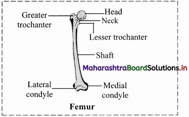
Explain the longest bone in human body.
Answer:
Femur: The thigh bone is the longest bone in the body. The head is joined to shaft at an angle by a short neck. It forms ball and socket joint with acetabulum cavity of coxal bone. The lower one third region of shaft is triangular flattened area called popliteal surface. Distal end has two condyles that articulate with tibia and fibula.
Internet my friend. (Textbook Page No. 212)
Question 1.
Find out information about types of fractures and how they heal.
Answer:
[Source: Tortora, G., Derrickson, B. Principles of Anatomy and Physiology. 15th Edition]
[Students are expected to find out more information about healing of fractures using the internet.]
Do you remember? (Textbook Page No. 208)
Question 1.
What are joints? What are the types?
Answer:
i. A point where two or more bones get articulated is called joint or articulation or athrosis.
They are classified based on degree of flexibility or movement they permit into lastly synovial or freely movable or diarthroses type of joints.
ii. Synarthroses / fibrous joints / movable joints:
In this joint, the articulating bones are held together by means of fibrous connective tissue. Bones do not exhibit movement. Hence, it is immovable or fixed type of joint. Synarthroses are further classified into sutures, syndesmoses and gomphoses.
iii. Cartilaginous / slightly movable joints / amphiarthroses:
These joints are neither fixed nor freely movable. Articulating bones are held together by hyaline or
fibrocartilages. They are further classified as
iv. Synovial joints / freely movable joints / diarthroses:
g. The types of synovial joints are on follows:
1. Pivot joint: In this type of joint, the rounded or pointed surface of one bone articulates with a ring formed partly by another bone and partly by the ligament. Rotation only around its own longitudinal axis is possible. e.g. in joint between atlas and axis vertebrae, head turns side ways to form ‘NO’ joint.
2. BaIl and socket joint: The ball like surface of one bone fits into cup like depression of another bone forming a movable joint. Multi-axial movements are possible. This type of joint allows movements along all three axes and in all directions. e.g. Shoulder and hip joint.
3. Hinge joint: In a hinge joint, convex surface of one bone fits into concave surface of another bone. In most hinge joints one bone remains stationary and other moves. The angular opening and closing motion (like hinge) is possible. In this joint only mono-axial movement takes place like flexion and extension. e.g. Elbow and knee joint.
4. Condyloid joint: It is an ellipsoid joint. The convex oval shaped projection of one bone fits into oval shaped depression in another bone. It is a biaxial joint because it permits movement along two axes viz, flexion, extension, abduction, adduction and circumduction is possible. e.g. Metacarpophalangeal joint.
5. Gliding joint: It is a planar joint, where the articulating surfaces of bones are flat or slightly curved. These joints are non-axial because the motion they allow does not occur along an axis or a plane. e.g. Intercarpal and intertarsal joints.
Saddle joint: This joint is a characteristic of Homo sapiens. Here the articular surface of one bone is saddle-shaped and that of other bone fits into saddle (each bone forming this joint have both concave and convex areas). It is a modified condyloid joint in which movement is somewhat more free. It is a biaxial joint that allows flexion, extension, abduction, adduction and circumduction. e.g. Carpometacarpellar joint between carpal (trapezium) and metacarpal of thumb.
Imagine. (Textbook Page No. 208)
Question 1.
If your elbow joint would be a fixed type of joint and joint between teeth and gum would be freely movable.
Answer:
Use your brain power. (Textbook Page No. 210)
Question 1.
Why are warming up rounds essential before regular exercise?
Answer:
Can you tell? (Textbook Page No. 211)
Question 1.
Classify various types of joints found in human body. Present the information in the form of chart. Give example of each type.
Answer:
i. A point where two or more bones get articulated is called joint or articulation or athrosis.
They are classified based on degree of flexibility or movement they permit into lastly synovial or freely movable or diarthroses type of joints.
ii. Synarthroses / fibrous joints / movable joints:
In this joint, the articulating bones are held together by means of fibrous connective tissue. Bones do not exhibit movement. Hence, it is immovable or fixed type of joint. Synarthroses are further classified into sutures, syndesmoses and gomphoses.
iii. Cartilaginous / slightly movable joints / amphiarthroses:
These joints are neither fixed nor freely movable. Articulating bones are held together by hyaline or
fibrocartilages. They are further classified as
iv. Synovial joints / freely movable joints / diarthroses:
g. The types of synovial joints are on follows:
1. Pivot joint: In this type of joint, the rounded or pointed surface of one bone articulates with a ring formed partly by another bone and partly by the ligament. Rotation only around its own longitudinal axis is possible. e.g. in joint between atlas and axis vertebrae, head turns side ways to form ‘NO’ joint.
2. BaIl and socket joint: The ball like surface of one bone fits into cup like depression of another bone forming a movable joint. Multi-axial movements are possible. This type of joint allows movements along all three axes and in all directions. e.g. Shoulder and hip joint.
3. Hinge joint: In a hinge joint, convex surface of one bone fits into concave surface of another bone. In most hinge joints one bone remains stationary and other moves. The angular opening and closing motion (like hinge) is possible. In this joint only mono-axial movement takes place like flexion and extension. e.g. Elbow and knee joint.
4. Condyloid joint: It is an ellipsoid joint. The convex oval shaped projection of one bone fits into oval shaped depression in another bone. It is a biaxial joint because it permits movement along two axes viz, flexion, extension, abduction, adduction and circumduction is possible. e.g. Metacarpophalangeal joint.
5. Gliding joint: It is a planar joint, where the articulating surfaces of bones are flat or slightly curved. These joints are non-axial because the motion they allow does not occur along an axis or a plane. e.g. Intercarpal and intertarsal joints.
Saddle joint: This joint is a characteristic of Homo sapiens. Here the articular surface of one bone is saddle-shaped and that of other bone fits into saddle (each bone forming this joint have both concave and convex areas). It is a modified condyloid joint in which movement is somewhat more free. It is a biaxial joint that allows flexion, extension, abduction, adduction and circumduction. e.g. Carpometacarpellar joint between carpal (trapezium) and metacarpal of thumb.
[Students are expected to prepare a chart on their own.]
Can you tell? (Textbook Page No. 211)
Question 1.
Human beings can hold an object in a better manner than monkeys. Why?
Answer:
[Note: Gorillas, chimpanzees, orangutans and some other variants of apes have opposable thumb.]
Internet my friend. (Textbook Page No. 211)
Question 92.
Now a days we hear from many elderly people that they are undergoing knee replacement surgery. Find out why one has to undergo knee replacement; how it is carried out and how it can be prevented.
Answer:
Knee replacement is done in following cases:
[Students are expected to find out more information about knee replacement on internet]
Find out. (Textbook Page No. 212)
Question 1.
You must have heard of Sachin Tendulkar suffering from ‘tennis elbow’, a cricketer suffering from a disorder named after another game. Can common people too suffer from this disorder? Find out more information about this disorder.
Answer:
[Students are expected to find more information about tennis elbow on their own.]